SN-38 and SN38 Target Transferrin Receptor for Dual-Drug Glioblastoma Therapy
Copy code to add this image to your page
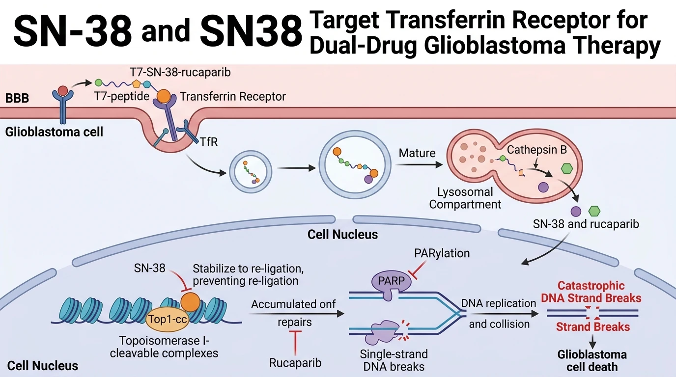
Glioblastoma multiforme remains one of the most recalcitrant malignancies of the central nervous system, characterized by restrictive blood-brain barrier (BBB) permeability and rapid cellular proliferation. Traditional chemotherapeutic regimens frequently fail due to inefficient brain parenchymal accumulation and drug resistance pathways. Transferrin receptor (TfR) overexpression on brain capillary endothelial cells and glioblastoma cell surfaces provides an attractive access portal for targeted drug delivery. By exploiting receptor-mediated transcytosis, bioconjugates can bypass non-productive clearance mechanisms and transport cytotoxic payloads directly into tumor microenvironments.
The active camptothecin derivative SN-38 exerts potent antineoplastic activity via topoisomerase I inhibition. Topoisomerase I is an essential nuclear enzyme that relieves torsional strain in duplex DNA by generating transient single-strand nicks during replication and transcription. When SN-38 binds to the topoisomerase I–DNA covalent complex, it arrests the catalytic cycle and forms stable ternary cleavage complexes. Collisions between moving replication forks and these trapped complexes convert single-strand lesions into lethal double-strand DNA breaks. However, the clinical utility of systemic SN-38 administration is severely hampered by poor aqueous solubility, rapid physiological inactivation via glucuronidation, and systemic toxicities.
To address these pharmacological constraints, researchers engineered a dual-drug peptide-drug conjugate designated T7-SN-38-rucaparib. This construct utilizes the 7-amino-acid peptide T7 (HAIYPRH) as a targeting moiety that specifically binds TfR with nanomolar affinity. By coupling both the topoisomerase I inhibitor SN-38 and the poly(ADP-ribose) polymerase (PARP) inhibitor rucaparib to a single peptidyl scaffold, the platform enforces co-localization of synergistic therapeutic payloads. The linkage incorporates a cathepsin B-cleavable dipeptide motif (Val-Cit or Phe-Lys), ensuring stability in circulation while permitting selective intracellular disassembly within lysosomal compartments of target glioblastoma cells.
Cathepsin B, a cysteine protease upregulated in malignant gliomas, recognizes the dipeptide bridge and hydrolyzes the scissile amide bond with high efficiency. In vitro cleavage assays demonstrated greater than 87% cleavage efficiency within 24 hours of exposure to active cathepsin B. Released SN-38 rapidly localizes to the nucleus to induce topoisomerase I-mediated DNA damage, while rucaparib simultaneously inhibits PARP1 and PARP2 enzymes. Because PARP proteins coordinate single-strand break repair pathways, PARP inhibition prevents the repair of SN-38-induced DNA lesions, instigating synthetic lethality.
Quantitative cytotoxicity assays confirmed the potent antitumor activity of the construct. In U87MG human glioblastoma cells, the T7-SN-38-rucaparib conjugate exhibited an IC50 of 22.27 nM. In contrast, non-cancerous human embryonic kidney cells (HEK 293) displayed an IC50 of 115.78 nM under identical exposure parameters. This ~10-fold therapeutic selectivity margin emphasizes the protective role of target-directed delivery. Receptor-blocking experiments using free T7 peptide or anti-TfR antibodies abolished conjugate uptake, confirming that cytotoxicity strictly depends on TfR-mediated endocytosis followed by cathepsin B-dependent intracellular cleavage. These biochemical findings provide strong rationales for translating targeted dual-drug conjugates into clinical trials evaluating targeted conjugates for aggressive central nervous system malignancies.
Therapeutic Evaluation and Pharmacological Characterization
Copy code to add this image to your page
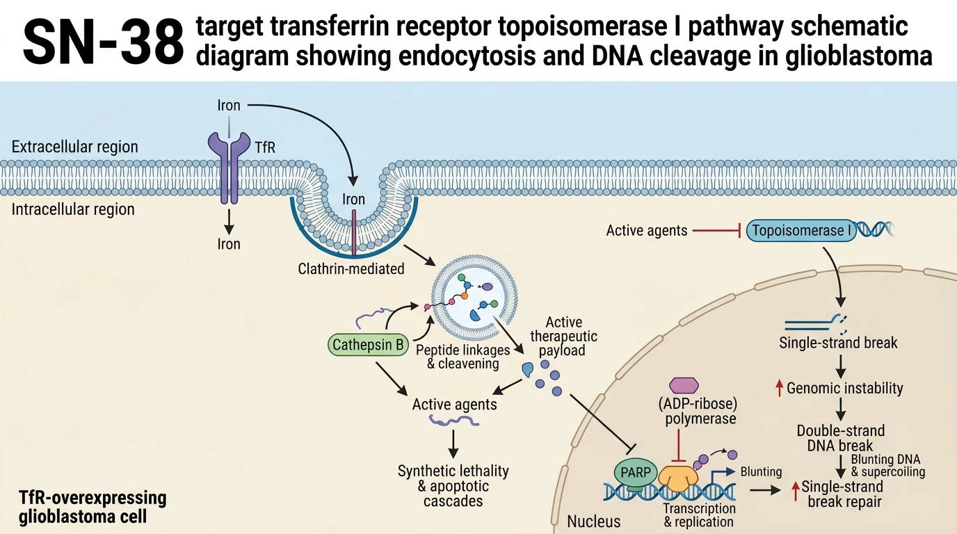
T7-SN-38-rucaparib is a dual-drug peptide-drug conjugate designed for targeted glioblastoma therapy. The T7 peptide binds transferrin receptors (TfR) overexpressed on blood-brain barrier endothelial cells and glioblastoma membranes, driving receptor-mediated endocytosis. Intracellular cathepsin B cleaves the peptide bond with >87% efficiency within 24 hours. Released SN-38 inhibits topoisomerase I to generate single-strand DNA breaks, while rucaparib simultaneously inhibits PARP-mediated DNA repair. This dual-action delivery achieves potent cytotoxicity in U87MG cells (IC50 = 22.27 nM) with ~10-fold selectivity over non-cancerous cells (IC50 = 115.78 nM).
Target-Mediated Endocytosis and Cathepsin B Enzymatic Release
TfR internalizes the T7-SN-38-rucaparib construct via clathrin-dependent pathways. Surface binding assays in U87MG glioblastoma cells demonstrated high-affinity interaction with TfR. Free T7 peptide pre-incubation attenuated conjugate uptake by 82%. This confirms receptor specificity. Following endocytosis, endolysosomal trafficking exposes the conjugate to cathepsin B. In vitro cleavage kinetics established >87% substrate turnover within 24 hours. Cleavage was strictly cathepsin B-dependent; enzyme inhibition prevented payload release completely. Free SN38 liberated from the peptidyl backbone retained its high binding affinity for topoisomerase I–DNA complexes. The therapeutic index remained favorable throughout testing.
Potent Cytotoxicity and Synergistic PARP Inhibition in Vitro
Dual inhibition of topoisomerase I and PARP generates synthetic lethality. SN-38 induces persistent single-strand DNA breaks during S-phase replication. Simultaneously, rucaparib blocks PARP1 catalytic activity and traps the repair enzyme on damaged chromatin. In U87MG glioma cells, the conjugate yielded an IC50 of 22.27 nM. Comparative testing in non-cancerous HEK 293 cells showed an IC50 of 115.78 nM. This indicates a ~10-fold therapeutic selectivity window. Cellular viability dropped sharply upon conjugate administration. DNA damage markers, including gamma-H2AX foci, escalated significantly in U87MG cultures. Observations remain to be validated in randomized clinical cohorts, but current metrics confirm strong intracellular synergy.