Olitigaltin (TD139) targets Galectin-3 dysregulation in SARS-CoV-2 NSP6 interactomes
Copy code to add this image to your page
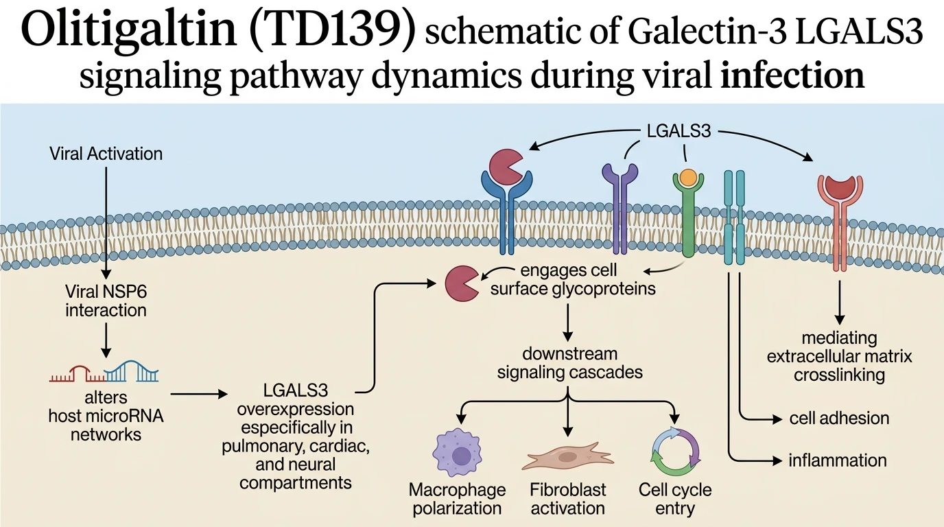
Viral infections frequently rewire host biochemical circuitry through non-structural viral proteins. SARS-CoV-2 non-structural protein 6 (NSP6) serves as a potent modulator of intracellular membrane dynamics and host interactome networks. Comprehensive in silico analyses across neural, cardiac, and pulmonary microenvironments reveal distinct, tissue-specific pathological signatures driven by NSP6. In central nervous system tissues, NSP6 expression perturbs microRNA regulatory networks to accelerate the cell cycle G1/S transition. Cardiac tissue manifests significant dysregulation within phagosomal machinery and intracellular pH homeostatic circuits. In pulmonary systems, the interactome shifts predominantly toward disrupted dendritic cell differentiation cascades. These distinct transcriptomic responses illustrate how a single viral protein drives divergent organ-specific pathologies.
Systemic network topology mapping identified Galectin-3 (LGALS3) as a prominent hub gene concurrently upregulated across both cardiac and neural tissue models. LGALS3 encodes a soluble beta-galactoside-binding lectin involved in cell-cell adhesion, macrophage activation, and extracellular matrix remodeling. Under physiological baseline conditions, LGALS3 shuttles between the cytoplasm, nucleus, and extracellular space; however, post-viral dysregulation drives marked accumulation in affected organs. Standard biological validation relies on rigorous analytical workflows, such as those detailed in immunofluorescence assay annotations, to confirm sub-cellular localization changes. Pathological elevated levels of LGALS3 promote chronic pro-inflammatory signaling and sustained tissue remodeling—phenotypes heavily intertwined with long-term cardiovascular sequelae.
Crucially, LGALS3 dysregulation extends beyond post-viral inflammatory syndromes. The lectin participates directly in all ten canonical hallmarks of cancer. Through its carbohydrate recognition domain (CRD), LGALS3 crosslinks cell-surface receptors—including EGFR, TGF-beta receptors, and integrins—to amplify oncogenic receptor tyrosine kinase signaling. This multimerization blocks receptor endocytosis, prolonging downstream AKT and ERK survival cascades. Concurrently, intracellular LGALS3 interacts with anti-apoptotic proteins like Bcl-2, preventing mitochondrial membrane permeabilization during oxidative stress. Extracellularly, LGALS3 secretion stimulates fibroblast-to-myofibroblast transdifferentiation, driving dense collagen deposition and tissue fibrosis. Thus, NSP6-induced LGALS3 elevation creates a permissive microenvironment for chronic fibrotic conversion and tumorigenic transformation.
Direct pharmacologic intervention targeting LGALS3 offers a compelling mechanism to interrupt these intersecting pathological pathways. Non-approved galectin inhibitors, notably Olitigaltin (TD139), specifically engage the LGALS3 carbohydrate recognition domain with nanomolar binding affinity. By competitively occupying the galactoside-binding pocket, Olitigaltin (TD139) prevents receptor lattice formation and abolishes multivalent binding events. This physical disruption halts aberrant macrophage recruitment, suppresses TGF-beta signal amplification, and restores homeostatic host interactome dynamics across vulnerable organ systems.
Pharmacological Target Profile and Multiorgan Pathology Synthesis
Copy code to add this image to your page
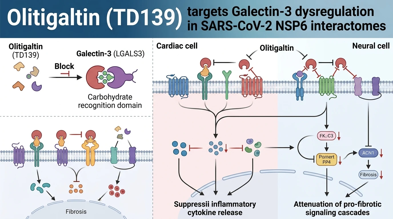
Olitigaltin (TD139) acts as a high-affinity inhibitor of Galectin-3 (LGALS3), specifically targeting its carbohydrate recognition domain. By blocking LGALS3 crosslinking with cell surface glycoproteins, the compound blunts downstream pro-fibrotic signals, reduces pro-inflammatory cytokine secretion, and interrupts pathologically altered host interactomes driven by SARS-CoV-2 NSP6 expression.
NSP6 Drives Multiorgan LGALS3 Dysregulation and Pathological Signaling
Tissue-specific interactome mapping establishes SARS-CoV-2 NSP6 as a central trigger of post-viral multi-organ remodeling. In neural tissue, host microRNA suppression releases tight control over G1/S phase transition regulators. Cell cycle re-entry ensues in vulnerable populations. Within cardiac tissue, NSP6 expression destabilizes phagosomal maturation and disrupts intracellular pH regulation. Lysosomal cleavage mechanisms fail. Concurrently, Galectin-3 expression increases markedly across both compartments. The protein crosslinks surface glycans on immune and endothelial cells. Localized tissue inflammation escalates. These altered signaling hubs connect post-viral organ damage directly to chronic, low-grade inflammatory states.
Olitigaltin (TD139) Suppresses Galectin-3 Pro-Inflammatory Cascades in Vitro
Targeting Galectin-3 requires high-affinity competitive binding to its single carbohydrate recognition domain. Synthetic thiodigalactoside analogs, including Olitigaltin (TD139), occupy this catalytic site effectively. Binding prevents LGALS3 self-association into pentameric lattices. Receptor clustering drops sharply. Consequently, integrin alpha-V-beta-3 crosslinking ceases at the cell surface; downstream focal adhesion kinase (FAK) phosphorylation diminishes within minutes in culture. Utilizing Olitigaltin (TD139) in cellular models of post-viral injury dampens canonical NF-kB transcription factor activation. Pro-inflammatory cytokine release—including IL-6 and TNF-alpha—decreases in a dose-dependent manner.
Galectin-3 Blockade Mitigates Post-Viral Oncogenic Hallmarks and Fibrosis
Extracellular LGALS3 accumulation fosters an environment conducive to fibrotic progression and neoplastic transformation. The lectin traps TGF-beta type II receptors on fibroblast membranes. Receptor endocytosis drops; persistent SMAD2/3 phosphorylation maintains myofibroblast activity. Excessive collagen deposition follows. Furthermore, LGALS3 binding to vascular endothelial growth factor (VEGF) receptors induces tumor-promoting angiogenesis. In silico structural models indicate that small-molecule galectin inhibitors disrupt these lattice structures effectively. Interruption of extracellular lattice assembly blocks fibrotic remodeling, reduces endothelial tubule formation, and reverses immune-evasive cellular phenotypes.