Dooku1 Mechanosensitive Cha Antagonism Accelerates Trigeminal Axonal Regeneration Post-Axotomy
Copy code to add this image to your page
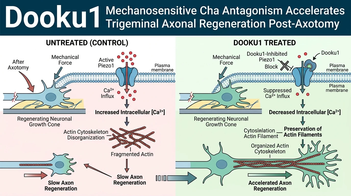
The mechanical microenvironment strictly governs peripheral nerve regeneration following traumatic injury. Mechanosensitive ion channels act as biological transducers, converting physical tension into intracellular signaling cascades that direct growth cone pathfinding. Among these mechanosensors, the pore-forming homotrimeric Piezo1 channel responds directly to lateral membrane stretch. Activation of Piezo1 allows rapid extracellular calcium influx, which triggers cytosolic calcium microdomains near the plasma membrane. In regenerating primary sensory neurons, uncontrolled calcium entry via Piezo1 engages calcium-dependent effectors, destabilizing the actin cytoskeleton and precipitating growth cone collapse. Consequently, basal or injury-induced mechanical stimulation acts as an intrinsic brake on neurite outgrowth.
Pharmacological inhibition of Piezo1 presents a targeted mechanism to bypass these mechanical inhibitory signals. The small molecule Dooku1 serves as an analog of the Piezo1 activator Yoda1, functioning instead as a selective Piezo1 antagonist. By occupying binding sites that modulate channel opening dynamics, Dooku1 blocks force-evoked channel gating without perturbing basal lipid membrane architecture. Evaluating the Dooku1 Mechanosensitive Cha antagonist profile requires advanced tissue culture architectures that accurately mimic the physical dynamics of nerve injury. Traditional static culture dishes fail to recapitulate the directional physical constraints and fluidic isolation required to evaluate isolated axonal injury and subsequent extension.
To rigorously assess axonal repair dynamics, modern experimental platforms utilize compartmentalized microfluidic chip models coupled with automated three-dimensional live-cell imaging. These microfluidic devices physically segregate the neuronal soma from the extending distal axons within microchannels. This configuration enables precise laser or fluidic axotomy while maintaining high-resolution longitudinal tracking of growing axonal tips across time. In primary trigeminal ganglion cultures subjected to fluidic axotomy, baseline spontaneous regrowth occurs slowly, restricted by local mechanical tension and intrinsic signaling repressors. Quantitative analysis of live-imaging series reveals that mechanical strain generated at the regenerating growth cone continually activates local Piezo1 complexes, maintaining an elevated baseline of local calcium transients.
Application of Dooku1 to injured trigeminal axons directly suppresses these local mechanotransductive braking signals. Dose-response evaluations demonstrate that Dooku1 administration enhances trigeminal axonal regrowth in a clear, concentration-dependent manner under microfluidic assay conditions. Quantitative live-cell imaging established that exposure to 30 µM Dooku1 produced a striking 154% increase in total axonal network length at 72 hours post-axotomy compared to untreated control channels undergoing spontaneous regrowth alone. At this concentration, Dooku1 treatment accelerated both the initial sprouting velocity of injured proximal stumps and the sustained directional elongation of growth cones across the microchannel interface.
Strikingly, the regenerative magnitude observed with 30 µM Dooku1 surpassed the pro-regenerative response elicited by standard neurotrophic factor combinations. Comparative cohorts treated with nerve growth factor combined with glial cell line-derived neurotrophic factor demonstrated significant but lesser network expansion over the same 72-hour window. While neurotrophin signaling primarily activates gene transcription and protein synthesis through receptor tyrosine kinase pathways, direct mechanochannel blockade directly prevents instantaneous calcium-mediated cytoskeletal destabilization at the immediate site of lesion. These empirical metrics confirm that relieving mechanical repression via Piezo1 channel antagonism yields superior elongation kinetics compared to traditional chemical growth factor stimulation in primary sensory neurons.
Collectively, these live-imaging observations delineate a crucial mechanistic role for Piezo1 channels in restricting peripheral axon repair. Uncoupling growth cones from local physical stress using Dooku1 transforms the mechanical injury interface into a permissive environment for rapid axonal re-extension. These data position Piezo1 channel blockade as a powerful biophysical therapeutic paradigm for nerve repair applications, particularly in corneal nerve injury and trigeminal neuropathies where physical microenvironments heavily dictate recovery outcomes.
Mechanistic Analysis of Piezo1 Antagonism in Axonal Repair
Copy code to add this image to your page
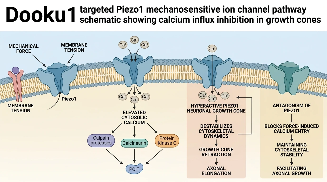
Dooku1 acts as a selective antagonist of the Piezo1 mechanosensitive ion channel. By blocking mechanical force-evoked channel opening, Dooku1 prevents intracellular calcium influx in damaged growth cones following nerve injury. Lowered cytosolic calcium levels preserve actin cytoskeletal dynamics, preventing growth cone collapse and overcoming intrinsic mechanical inhibition of axon growth. In trigeminal nerve axotomy models, this mechanochannel inhibition drives dose-dependent axonal network regrowth, outperforming classical growth factor stimulation.
Piezo1 Suppression Prevents Calcium-Induced Growth Cone Collapse
Mechanical tension across damaged axonal membranes rapidly opens Piezo1 ion channels. This activation triggers localized intracellular calcium surges near the growth cone tip. High calcium levels activate calpain proteases; subsequent cleavage of cytoskeletal anchor proteins causes immediate growth cone collapse. Pharmacological application of the selective Dooku1 Mechanosensitive Cha inhibitor suppresses these force-dependent calcium influx events. In microfluidic trigeminal axotomy assays, blocking Piezo1 maintains f-actin bundle organization during growth cone extension. Cytoskeletal stability remained intact throughout the 72-hour observation period. Intracellular calcium fluxes dropped significantly upon target engagement. Eliminating local calcium spikes preserves growth cone morphology, allowing continuous filopodial extension across microchannels despite physical substrate constraints.
Dooku1 Outperforms Dual Neurotrophin Stimulation in Axotomy Models
Standard therapeutic paradigms for peripheral nerve damage rely on exogenously added growth factors. However, combined administration of NGF and GDNF yielded inferior total axonal network lengths compared to single-agent treatment with 30 µM Dooku1. Quantitative live-cell tracking demonstrated a 154% increase in network length at 72 hours post-injury. Mechanical inhibition appears to represent a primary rate-limiting barrier to sensory nerve extension. Releasing this mechanical brake allows rapid growth cone advance without requiring hyper-activated transcriptional cascades. Under tested microfluidic culture conditions, direct channel blockade provided superior kinetic responses. These experimental metrics indicate that Piezo1 channel antagonism effectively circumvents intrinsic mechanical repressors in regenerating trigeminal axons.
Therapeutic Implications for Corneal and Trigeminal Nerve Repair
Corneal nerve damage often leads to persistent sensory deficits and impaired epithelial healing. The trigeminal ganglion directly innervates corneal tissue, making trigeminal axotomy models highly relevant for ocular nerve pathologies. Within this in vitro model system, targeted modulation of Piezo1 mechanoreceptors demonstrated substantial axonal recovery kinetics. Suppressing mechanosensitive channels allows growth cones to navigate surrounding dense extracellular matrices without undergoing arrest. While these observations remain to be validated in vivo, the clear dose-dependent response establishes a strong pharmacodynamic rationale. Dooku1 represents an important lead molecule for developing topically applied therapeutics aimed at corneal nerve regeneration.