BMS-202 PD-1/PD-L1 Checkpoint Inhibition via Radiopaque Bead Delivery for Hepatocellular Carcinoma
Copy code to add this image to your page
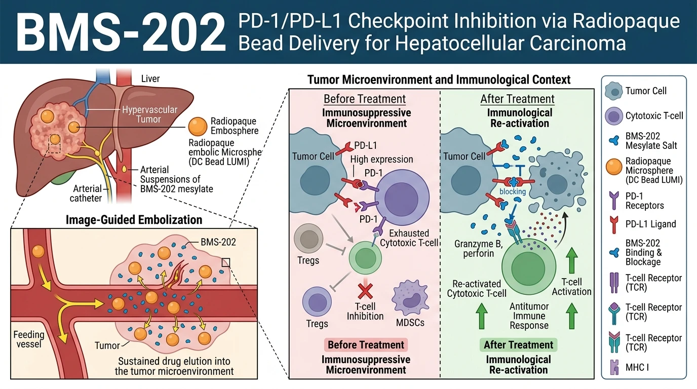
Immune checkpoint blockade targeting programmed cell death protein 1 (PD-1) and its primary ligand (PD-L1) has transformed clinical oncology. However, monoclonal antibodies face distinct pharmacokinetic limitations in solid tumors. These include poor tissue penetration, prolonged systemic half-lives, and immune-related adverse events. Hepatocellular carcinoma (HCC) presents an additional hurdle due to dense tumor stroma and altered vascular architecture. Locoregional delivery strategies offer a targeted alternative. By administering therapeutic agents directly through tumor-feeding arteries, high local concentrations are attained while reducing systemic toxicity. The small molecule BMS-202 provides a synthetic solution to checkpoint inhibition by inducing PD-L1 dimerization and sterically blocking PD-1 interaction.
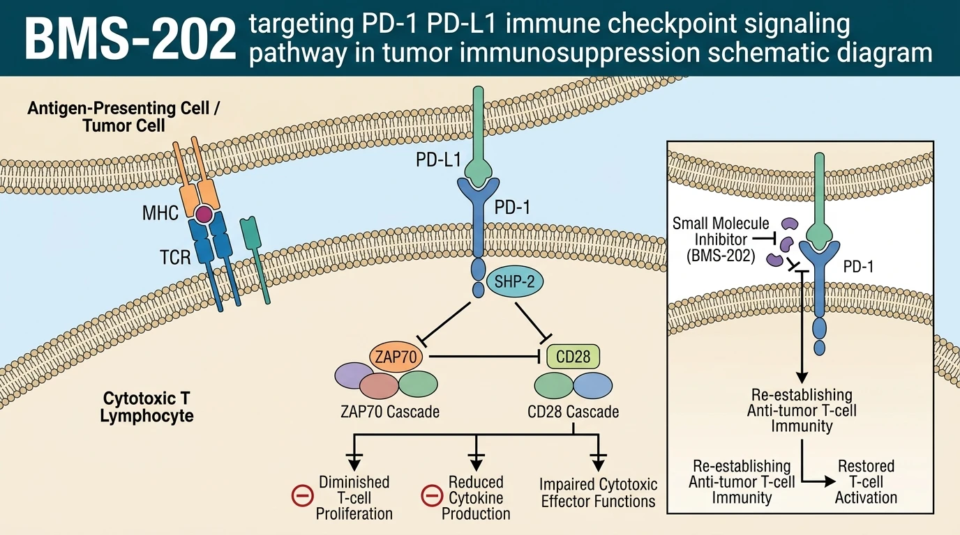
Traditional monoclonal antibodies bind the flexible extracellular domain of PD-L1 with high affinity. BMS-202 operates through a distinct biophysical mechanism. It binds directly to homodimeric interface pockets of PD-L1. This structural rearrangement sequesters the receptor-binding surface. Consequently, interaction with cell-surface PD-1 on CD8+ T cells is prevented. Converting BMS-202 into a mesylate salt improves aqueous solubility, enabling incorporation into ionic hydrogel delivery systems. Standard transcatheter arterial chemoembolization (TACE) relies on cytotoxic drugs like doxorubicin loaded into hydrogel microspheres. Adapting this platform for small-molecule immunomodulators requires precise chemical pairing between drug species and bead matrix chemistry.
Radiopaque embolic beads, such as DC Bead LUMI, feature sulfonate-modified polyvinyl alcohol (PVA) hydrogel networks impregnated with triiodobenzene moieties. These sulfonate groups facilitate ionic binding with positively charged basic molecules. In quantitative evaluation, BMS-202 mesylate displayed drug loading capacities reaching 148.05 ± 2.24 mg/mL of hydrated beads. This value exceeds the theoretical ionic exchange capacity of the bead matrix by approximately 23%. This secondary capacity suggests that loading depends on more than simple electrostatic interaction. Non-covalent hydrophobic interactions between the aromatic ring structures of BMS-202 and the hydrophobic segments of the PVA polymer hydrogel drive additional matrix partitioning. This dual loading mechanism allows high-density payload retention within the radiopaque microspheres.
Elution kinetics govern the local therapeutic window during transcatheter procedures. Rapid washout reduces local residency time, rendering locoregional delivery ineffective. Under controlled in vitro wash conditions, BMS-202 showed minimal premature leakage during initial aqueous preparation steps. When introduced into physiological dissolution media, the molecule exhibited a sustained, controlled elution profile similar to clinical doxorubicin standards. Drug retention remained stable without immediate burst release. These kinetic properties indicate that BMS-202 can be locally delivered using standard interventional radiology protocols. The integration of small-molecule immune checkpoint inhibitors into radiopaque beads creates opportunities for image-guided immuno-chemoembolization. Further research using chemical biology research platforms will clarify how local drug depots alter intratumoral T-cell exhaustion and myeloid cell polarization in primary liver tumors.
Therapeutic Evaluation and Delivery Kinetics in Hepatocellular Carcinoma
Copy code to add this image to your page
BMS-202 acts as a small-molecule inhibitor of the PD-1/PD-L1 interaction by inducing PD-L1 homodimerization and blocking receptor engagement. Formulated as a mesylate salt, BMS-202 achieves robust loading (148.05 ± 2.24 mg/mL) into radiopaque hydrogel microspheres via combined ionic and hydrophobic interactions. The loaded microspheres demonstrate controlled, sustained release without rapid initial washout, matching clinical doxorubicin elution kinetics. This system provides a strategy for image-guided immuno-chemoembolization in hepatocellular carcinoma models.
Supramolecular Loading Exceeds Theoretical Ionic Exchange Capacity
Quantitative binding assays confirmed that BMS-202 mesylate interacts efficiently with sulfonate-functionalized PVA hydrogel matrices. Electrostatic attraction between the protonated amine functions of the drug salt and the negative sulfonate centers initiates bead loading. Empirical measurement demonstrated a maximal binding capacity of 148.05 ± 2.24 mg/mL of hydrated beads. This figure exceeded theoretical ionic exchange thresholds by ~23%. Secondary non-ionic forces account for this supramolecular assembly. Aromatic pi-stacking and hydrophobic interactions between BMS-202 molecules and the polymer backbone facilitate localized self-aggregation within the hydrogel pores. Within this specific hydrogel context, the dual binding modality enables dense drug loading without causing physical bead deformation or structural aggregation during preparation.
Sustained Hydrogel Elution Prevents Rapid Hydrodynamic Washout
Sustained local release remains essential for localized checkpoint blockade within hypervascular hepatocellular carcinoma lesions. In dissolution testing, BMS-202 showed stable matrix retention during routine handling and aqueous processing. Immediate burst release did not occur upon exposure to fluid currents. The drug eluted in a controlled fashion over extended periods. This kinetic profile mirrors established doxorubicin release dynamics from similar PVA-based platforms. Small-molecule BMS-202 PD-1/PD-L1 disruption requires sustained microenvironmental drug concentrations to maintain T-cell receptor signaling across surrounding tumor tissue. These preliminary data support the feasibility of image-guided immuno-chemoembolization using radiopaque microspheres. Observations remain to be validated in hypervascular animal models and randomized clinical cohorts.